LUMBAR SPINE REHABILITATION
INTRODUCTION & anatoMY
Lower back pain is a very common condition that affects many individuals across a variety of age groups, occupations and athletic pursuits. Sometimes it relates to an acute injury or event, and other times it may gradually build over time. In some cases, it can even be a chronic condition. Regardless of the cause, most lower back pain resolves within a few months with appropriate rehabilitation, and its bark is usually worse than its bite. Having a good understanding of the lower back, its anatomy and what your pain means will help you to progress and make a good recovery. A good mechanic would never try to fix a car without first understanding its parts, how they function and the nature of the problem. Your back is no different!
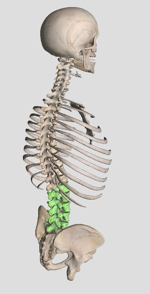
The lower back ('Lumbar Spine') refers to the last 5 vertebrae in our spine, ending at our tailbone.
Complete Anatomy Version 2.3.7 (4120), 2017 (1)
ANATOMY OF THE LOWER BACK (LUMBAR SPINE)
The lower back - called the 'lumbar spine' - refers to the lower most part of our spine that finishes at our tailbone (sacrum) and hips. It comprises 5 lumbar vertebrae, spaced apart by intervertebral discs, and connected by facet joints and ligaments. Inside the vertebral arch lies the spinal cord and lower spinal nerves, and nerve roots exit between each of the vertebrae before travelling down to the legs to provide sensation and contraction of our muscles.
Complete Anatomy Version 2.3.7 (4120) (2017) (1)
The spine is composed of vertebrae, spaced apart by intervertebral discs, and connected together by facet joints and ligaments. Spinal cord tissue is enclosed in the vertebral arch. Nerve roots exit in the spaces between vertebrae. The lower back - 'lumbar spine' - refers to the 5 lowest vertebrae just before the tail bone and hips.
The lower back is a beautifully complex and inherently robust structure. The vertebrae themselves are very large and designed for weight bearing, and the discs and ligaments are very strong, binding the vertebrae together. The discs have a nucleus (inner jelly like paste - shown in blue in the above diagram) and hard outer rings of collagen called the annulus (shown in white in the above diagram). Each of the facet joints that connects vertebrae from above and below has cartilage, a joint capsule and joint fluid, just like any other joint in our body. These structures give passive stability to the spine, hold it together tightly and guide its movement.
Back view of deep muscles
Complete Anatomy Version 2.3.7 (4120) (2017) (1)
Front view of deep muscles
The spine has multiple layers of muscle. Deeper muscle layers (above) help to bind adjacent vertebrae together, providing reactive stability to the spine and adjustment of tension. Larger overlying muscles (below) help to generate movement and force. This allows the spine to be both robust and mobile.
Back view of superficial muscles
Complete Anatomy Version 2.3.7 (4120) (2017) (1)
Side view showing abdominal muscle wall
Like cables holding down a radio tower, the spine is also supported by a network of muscles that provide reactive support (adjustment of stiffness and tension) to control load and generate movement. Some of these muscles (such as multifidus, psoas and quadratus lumborum) connect one vertebrae to another and help to ensure the segments of the spine are stabilised. Other muscles are much larger and span across multiple vertebrae (such as the erector spinae and abdominal muscles), helping to generate force and movement, and couple movement of the lower back to that of our hips. Like a pivot point under a crow-bar, the small muscles allow the large muscle to generate force in a stable fashion, ensure even loading and leverage. Abdominal muscles in conjunction with the diaphragm and pelvic floor muscles also help to generate abdominal pressure (like an internal balloon of air), increase stiffness and stability of the spine and control rotational movements. Of particular note is the deep transversus abdominus muscle that acts like a muscular corset or built in weightlifting belt.
Complete Anatomy Version 2.3.7 (4120) (2017) (1)
The diaphragm and pelvic floor muscles (left) in conjunction with the deep transversus abdominus muscle (right) form muscular walls around the abdominal cavity, helping to stabilise and stiffen the lumbar spine when they contract.
The multiple layers of muscle surrounding the spine and abdomen are held together by connective tissue that acts like a sort of glue - binding all layers and transmitting forces. This connective tissue is known as fascia. Fascia adds further stability to the spine and in the lower back there is a thickened superficial layer of fascia - known as the thoracolumbar fascia. This structure adds further strength and control in this region of the spine. In addition to the tissues directly attached to the spine, the tissues associated with the pelvis and hips are important to the function of the lumbar spine. Of particular note are the hip flexor, gluteal and groin muscles. By their attachment to the lumbar and abdominal fascia, pelvis and legs, these muscles are very important for providing a stable base of support and adjusting posture of the lumbar spine. In a similar fashion, the large latissimus dorsi muscles in the upper back/shoulder can help to stabilise the lumbar spine by bracing the facial tissue when they contract.
Complete Anatomy Version 2.3.7 (4120) (2017) (1)
The latissimus dorsi and gluteal mucles attach to the thoracolumbar facia and help to brace the spine. Similarly, the groin and hip flexor muscles are associated with the abdominal muscles and fascia and can help to control the spine.
In viewing the anatomy of the lumbar spine its complexity can be appreciated. Furthermore, so can its strength. With the synchrony between the ligaments, discs, joints, supporting muscles and fascia, the spine is incredibly strong. Indeed, biomechanical studies of weightlifters' lumbar spines suggest that they tolerate as much as 17,192 N of compressive load (1,753 kg!) when lifting heavy weights (2). Of course these are incredible feats of strength that weightlifters can perform with the right training, but it goes without saying that when it is working optimally the human spine is tough!
1. 3D4Medical Ltd. (2017).Complete Anatomy (Version 2.3.7 (4120)) [Windows Application Software]. Retrieved from http://3d4medical.com
2. Chollewicki, J. McGill, SM. Norman, RW. (1991). Lumbar spine loads during the lifting of extremely heavy weights. Medicine and science in sports and exercise, 23(10), 1179-1186.


